Showing posts with label Sports Medicine Acupuncture. Show all posts

Wednesday, December 13, 2017

Iliac Crest Syndrome

Iliac Crest Syndrome - A Common Cause of Low Back Pain

Fig. 1: Pain site at the posterior iliac crest region

Pain experienced at the iliac crest is a frequent low back condition affecting patients seeking help from Sports Medicine Acupuncturists®. The iliac crest is the top (or ‘crest’) of the ilium, which is the most superior or upper portion of the pelvic structure. If you were to place your hands on your hips, they would be resting on the iliac crest. Pain at the iliac crest, referred to as ‘iliac crest syndrome,’ is experienced at the posterior (back) portion of the iliac crest and can be persistent (Fig. 1).

Fig. 2: Palpation of yaoyan at 
the superficial and deep vectors
This pain is at an acupuncture point called yaoyan which is a commonly used 'extra point'. Extra points are points which are not on main acupuncture channels, but have been found to be clinically important nonetheless. This particular extra point is found at the attachment site of two important back muscles. Depending on the depth, these muscles are either the iliocostalis lumborum or the quadratus lumborum (Fig. 2).

The iliocostalis lumborum is the more superficial of the two of these muscles. It is one of three muscles which are part of a group called the erector spinae (Fig 3 left image). This is the group of muscles that span the back from the hip through the neck and run parallel to the spine. The iliocostalis lumborum is the ‘lumbar’ or low back portion of this group; it runs from the top of the iliac crest (the ‘ilio’ part of the name) to the ribs (the ‘costo’ part of the name). This muscle then continues upward (but it is then called the iliocostalis thoracic and iliocostalis cervicis) and is the most lateral of the three muscles of the erector spinae. The iliocostalis functions with the other muscles of this group to perform extension of the torso, which is the motion involved in bending backward. However, since this muscle is a bit more lateral than the others in the group, it is also involved in side bending motion. In this case, only one side is primarily involvedthe right muscle in right side bending and the left in left side bending.

The quadratus lumborum is a deeper muscle underneath the iliocostalis (Fig. 3 right image). It runs from the iliac crest and has attachments on the lumbar (low back) vertebrae, and ends at the 12th (lowest) rib in the back. This muscle laterally flexes the trunk. It does this by shortening the space between the top of the hip and the 12th rib. This would either pull the rib towards the hip (sidebending on that side) or pull the hip towards the rib (elevating or hiking the hip up on that side).

Both of these muscles have attachments on the iliac crest and both can become pain-producing sites. In both cases, these muscles would be in a shortened position when the hip is hiked on the side of pain. This is frequently what is seen with iliac crest pain.
Fig. 3: Image on the left shows the iliocostalis which is the lateral muscles of the erector spinae group. The image on the right has this group removed to highlight the deeper quadratus lumborum muscle. Both are common sites of pain at the iliac crest. Both images are from Netter's Atlas of Human Anatomy.
When the hip is elevated on one side, as is often the case with iliac crest syndrome, it is not simply the muscles discussed which are involved. Other muscles whose job it is to stabilize the hip and prevent it from elevating are also part of the overall picture. The gluteus medius and minimus are the primary muscles which do this, and these muscles have a propensity to become inhibited and fail in their stabilization roll.


When treating iliac crest syndrome, it is important to address all of the muscles involved in the imbalance. This includes both the shortened and overactive muscles such as the iliocostalis and quadratus lumborum, along with the inhibited and overlengthened muscles such as the gluteals. Acupuncture and manual therapy are powerful treatment options to correct these imbalances, and corrective exercises performed by the patient can solidify treatment at continue to return function.


Facebook icon Google Search icon LinkedIn icon Instagram icon YouTube icon

Thursday, March 2, 2017

Injures Caused by Foot Overpronation

Foot Overpronation Can Cause Many Different Injures.

Pronation of the foot involves dorsiflexion of the ankle, abduction of the foot, and eversion of the foot. In layman's terms this is described below, but the important aspect is that these movements drop the medial arch of the foot.
  • Dorsiflexion involves a bend of the ankle in the direction of the dorsum or top of the foot. It brings the top of the foot towards the knee.
  • Abduction is a turning out of the foot away from the midline.
  • Eversion is a turning out of the foot which brings the medial arch down.
Fig. 1: Comparing a neutral foot (middle) to foot overpronation (left) and supination (right). Notice how the pronated foot turns away from the midline (this is abduction) and the medial arch falls towards the floor (this is eversion). Image Ducky2315 (Own work) [CC BY-SA 3.0 9http://creativecommons.org/licenses/by-sa/3.0)]

A certain amount of pronation is normal in weight bearing as this acts as a shock absorption. Connective tissue structures in the foot elongate and produce a certain amount of tension which, upon recoil, helps propel the movement, thus acting as an energy saving mechanism during walking and running. However, overpronation is a frequent occurrence which is an excessive pronation. This can lead to a range of injuries of the foot, ankle and lower leg, but also can cause or contribute to injuries of the knee, hip and really anywhere in the body.

Frequent injuries are listed below with a brief description of how overpronation contributes.

  • Plantar fasciitis/fasciosis: As the foot goes excessively into pronation, the plantar fascia is overstretched. The reoccurring motion eventually irritates the plantar fascia and can lead to chronic pain in the bottom of the foot, usually where this tissue attaches to the calcaneus.
Fig. 2: Both tibialis anterior and tibialis posterior attach to 
the medial arch. In foot overpronation, the arch drops and these 
muscles are pulled long. This can irritate the tendons of these 
muscles and can also contribute to shin splints.

  • Tibialis posterior tendinopathy: This condition can be misdiagnosed as plantar fasciitis, but pain is usually more at the medial ankle. The tibialis posterior supports the medial arch and can become irritated with excessive pronation as it too is repetitively overlengthened when walking or running.
  • Shin splints: Shin splints can be classified as anterior or medial shin splints. The involve either the tibialis anterior or the tibialis posterior, respectively. Both of these muscles support the medial arch and can have a shearing effect where they attach to the tibia with overpronation.
Fig. 3: Note the effects on the knee and hip.
  • Knee pain: There are many knee conditions that can become aggravated with overpronation. As the foot pronates, the lower leg turns inward. This becomes excessive with overpronation, and the knee tends to collapse in. This can contribute to medial knee pain from conditions such as pes anserine tendinopathy or lateral knee pain from conditions such as iliotibial band friction syndrome.
  • Hip pain: Like knee pain, there are many hip conditions that can be aggravated from foot overpronation. One example is greater trochanteric bursitis which is often caused by an excessive raising of the hip during weight bearing. This frequently occurs with foot overpronation and with the knee moving in.
  • Back pain, shoulder pain, neck pain: Foot overpronation can be involved with many other muscle imbalances as described above with knee pain and hip pain. These imbalances can affect regions as far away as the neck.
Sports Medicine Acupuncture® is a great system for treating both the injury and the underlying causes of the injury such as foot overpronation. It employs acupuncture, manual therapy, and corrective exercises in addition to other possible treatment options. Both local acupuncture and manual therapy techniques can be used to reduce pain and improve the health of the injured soft tissue. And acupuncture to specific points within the muscles can correct the underlying muscle imbalances that occur with foot overpronation which lead to the injury and, if uncorrected, will cause a recurrence. Corrective exercises help the treatment hold and further corrects the underlying muscle imbalance.


Fig. 4
A simple exercise which can strengthen the intrinsic muscles of the foot and help to correct foot overpronation is the short foot exercise. This exercise strengthens the adductor hallucis (hallucis refers to the big toe) muscle (Fig. 4), in particular. The following steps are followed to perform this exercise:


  1. Sit upright with both feet flat on the floor. Alternately, the exercise can be performed standing on the foot to be exercised. This is more advanced.
  2. Raise the arch of your foot by bringing your big toe towards your heel. The trick is to do this without curling your toes. Your emphasis is on lifting the arch away from the floor.
  3. Hold for 5-10 seconds. You can perform this exercise multiple times. 
While this exercise is helpful, foot overpronation often involves other structures not only in the foot. These include muscle imbalance in the lower leg, knee and thigh, hip and low back. Comprehensive work with these structures is often necessary for lasting effects. Evaluation and treatment of these contributing muscle imbalances along with treatment of associated injuries are what your certified Sports Medicine Acupuncturists® work with.


Facebook icon Google Search icon LinkedIn icon Instagram icon YouTube icon

Tuesday, August 23, 2016

High Hamstring Tendinopathy

High Hamstring Strain and High Hamstring Tendinopathy

Fig 1: Common mechanism of
hamstring injury
High hamstring strain and/or pain at the attachment of the hamstrings (referred to as high hamstring tendinopathy or proximal hamstring tendinopathy) is a common complaint among runners and athletes who are involved in kicking sports such as soccer. The pain is experienced at the region of the sit bones (the ischial tuberosity) and is aggravated with sitting and with activities such as running and kicking. The pain can be sharp, aching, and sore, and it can be described as a pulling sensation. This injury can be very slow to heal and has a high frequency of recurrence.

The hamstrings frequently become injured in their eccentric phase of contraction. What this means is that in activities such as kicking a soccer ball or football, or in the leg swing during running, the hamstrings are lengthening (and the quadriceps are contracting and shortening). Although the hamstrings are lengthening, they fire to slow down or decelerate the motion (Fig.1).

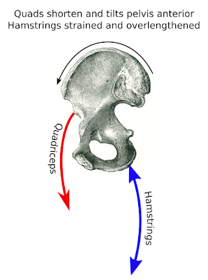
Understanding this mechanism of injury is crucial for Sports Medicine Acupuncturists® such as myself, as this informs clinical inquiry. Are the quadriceps and other hip flexors short and overactive placing the hamstrings in a chronically taut and lengthened position (Fig. 2)? This would need to be addressed to allow adequate healing. Or, are the hamstrings chronically in a shortened position? Both of these situations could be an underlying cause of hamstring strain recurrence and in both situations the patient would complain of 'tight' hamstrings. In the first instance these 'tight' hamstrings are pulled taut like a rubber band overstretched (a very common occurrence), while in the second they are in a short and tight positions. However, treatment strategies for these two instances would be very different.

Fig. 2: Schematic illustrating hamstrings being pulled in a chronically overlengthened position. Patients would complain of chronically 'tight' hamstrings in this case as they are chronically pulled taut. 


Fig. 3: Modified from Netter's
Atlas of Human Anatomy
When patients complain of this pain, it is also important to properly assess which structure is actually injured. While many patients come in to clinic complaining of high hamstring strain, two other structures frequently mimic this pain. The lower fibers of the gluteus maximus overlie the hamstrings at the region where high hamstring strain occurs. When this muscle is the culprit, there is a palpable taut band that can be felt in these fibers from about the region of the coccyx to the attachment of the hamstrings. Also, the adductor magnus, the most posterior muscle in the adductor group which is on the medial thigh, attaches very close to the hamstrings and pain associated with this muscle can mimic high hamstring strain (Fig. 3). Both of these muscles create a slightly different 'flavor' of pain and can have characteristic signs and symptoms reported by the patient; this allows an astute clinician to find the fixed site of pain and treat it properly for the quickest healing time.


Besides these sources of pain, referred pain is also a possibility and needs to be considered. Referred pain may come from the lumbar spine, from the sacroiliac joint, or from trigger points in the gluteal muscles, the low back, or even the lower portion of the hamstrings. The clinician must be thorough during evaluation and patients should make sure that their practitioner, whether an MD, acupuncture physician, physical therapist, or massage therapist, has the understanding and training to properly assess and evaluate the condition. Proper assessment of all the factors leads to proper treatment. Proper treatment leads to faster and more profound healing.


Facebook icon Google Search icon LinkedIn icon Instagram icon YouTube icon


Facebook icon Google Search icon LinkedIn icon Instagram icon YouTube icon

Saturday, August 6, 2016

Levator Scapula Neck Pain

Levator Scapula Disfunction Can Cause Pain and Stiff Neck Syndrome

Next week I will be teaching a class at the Florida State Oriental Medical Association (FSOMA) Annual Conference. The class will discuss neck pain and stiffness and will specifically highlight acupuncture and manual therapy techniques to treat the levator scapula, a muscle which frequently causes neck pain and stiffness and pain in the shoulder blade region.

Fig. 1 A (left) and 1 B (right): Images from Myofascial Pain and Dysfunction: The Trigger Point Manual, by Drs. Janet Travell and David Simons,

The levator scapula, seen in Fig. 1 A, is a muscle which stretches from the upper neck to the upper part of the shoulder blade. Not surprisingly based on its name, the levator scapula elevates the scapula. But it also rotates bends it sideways. Both of these movements basically move the shoulder blade closer to the neck on the same side. When this muscle develops trigger points (painful nodules within the muscle), it can cause quite a lot of pain and neck stiffness. This pain pattern is illustrated in the Fig. 1B and as can be seen, the pain concentrates at the base of the neck and frequently spreads to the medial border of the scapula. Patients often state that they feel the pain between the shoulder blades. The neck frequently becomes stiff, and pain is worse when patients turn the head to the side of the pain, as in looking over the shoulder.


Fig. 2: Neck position which shortens the levator scapula and can lead to pain.


Fig. 3: The imbalance in the pelvis
and legs, causing an elevation on
of the right ilium which frequently
contributes to an elevation of the
left shoulder girdle. This can
cause pain and stiffness in the
left neck.
This can be a chronic condition, or it can come on suddenly. It is not uncommon for people to wake with this pain after a night's sleep in an awkward position. A cold draft on the neck while sleeping is also frequently reported by patients. This pain can be quite distressing as it interferes with activities such as driving and makes it difficult to find a comfortable position.

In chronic cases, it is found that patients often perform an activity that repetitively shortens this muscle. A common example is a busy office worker, student or parent holding a phone to their ear with their shoulder. Awkward computer workstations, poor sitting posture, poor breathing, and even imbalances affecting the leg length (more on this in a different post) can contribute to pain in the levator scapula (Fig. 3)


Local acupuncture techniques can be an excellent way to address the muscle directly to release muscle contraction. In addition, acupuncture along the related channels and to muscles that are part of the dysfunction, myofascial release (a deep tissue type of massage) to lengthen bound muscles and connective tissue, and corrective exercises to address posture are all tools that can greatly reduce pain and improve range of motion in the neck and treat this 'stiff neck syndrome.'

A self-help exercise is described below. This can be performed several times a day and should not cause pain or aggravation of symptoms. Use your judgment and consult your physician if you have any doubts.

This exercise is described as if there is pain at the base of the neck on the left side, which is worse when turning to the left. The directions can be reversed for pain occurring on the right side.

1) Lie face-up or sit upright in a chair with your feet on the floor.
2) Gently turn your neck towards the painful side (to the left) to the point just before it hurts (this may be only a small turn in severe cases).
3) Place the hand opposite the painful side on your cheek (right hand on the right cheek) and gently, with very little force, turn the head back into the palm (to the right). There should be no movement, and this is an isometric contraction. In other words, you are resisting the gentle turn with your palm and not allowing any movement. Hold this position for about 6 seconds.
4) Relax for about 1 second, and then see if you can turn more towards the painful side (to the left). Still stop before there is actual pain and do not attempt to turn more than your body will allow.
5) Repeat steps 3 and 4.

Note: This exercise is most useful in acute problems when there is severe pain and difficulty turning the neck. The goal is to GENTLY tease out movement. Many times, the body perceives that there is danger to the joints (maybe you fell asleep in a position that was stressing the joints of the neck, for instance) and there is a reflexive spasm to guard and prevent movement. Trying to stretch aggressively and forcefully will often aggravate the condition more in these situations, as the neck muscles such as the levator scapula will contract more to guard the area. In more chronic cases, stretching and range of motion exercises can be employed.


Facebook icon Google Search icon LinkedIn icon Instagram icon YouTube icon

Tuesday, January 19, 2016

Frozen Shoulder and Related Conditions

Supraspinatus tendinopathy, bicipital tenosynovitis, infraspinatus myostrain, and subscapularis myostrain can often be diagnosed as Frozen Shoulder.


“Frozen shoulder” is a common diagnosis given for those experiencing shoulder pain, stiffness and a decrease in mobility. True frozen shoulder, which is called adhesive capsulitis, refers to a condition where there is a buildup of scar tissue in the joint capsule of the shoulder. There exist several other conditions which often get called “frozen shoulder,” but those can be more precisely differentiated. The most common of these conditions that I see in clinical practice are: supraspinatus tendinopathy; bicipital tenosynovitis; infraspinatus myostrain; and subscapularis myostrain. Each of these involves different structures, has variation in pain patterns and referrals, and, more importantly, requires a different treatment for a successful outcome.
Frozen shoulder and related conditions affect many people of different ages and they affect both men and women. Those between 40-60 years of age are more frequently afflicted with shoulder problems, and women experience shoulder pain much more frequently than men. 2-5% of the population in this age demographic will experience this problem. There is evidence showing that hormonal changes during menopause are linked to the greater frequency of shoulder pain experienced by women.
These conditions may be caused by trauma or they may come on insidiously, in which case there seems to be no apparent cause, although it’s possible that postural imbalances, age, nutritional deficiencies, changes in hormone levels, diabetes, Parkinson’s and other conditions may be contributing factors. Proper differentiation is crucial when treating so that mechanism of injury can be determined, target tissue can be assessed and treated, postural disparities can be corrected, and proper use of medicinals can be employed (herbals, nutraceuticals, or pharmaceuticals depending on the scope and treatment philosophy of the physician). In Sports Medicine Acupuncture®, this is extremely important as the injury or cause of pain will guide the proper use of acupuncture, myofascial release (a type of clinical, deep tissue massage), corrective exercises, and herbal medicine prescription.
I will discuss the four main causes of shoulder pain and stiffness in future posts and will link these blog posts together. Again, these conditions are:
  • Supraspinatus tendinopathy – an inflammation or irritation with fibrosis (extra fibrous tissue) of the tendon of the supraspinatus (one of the rotator cuff muscles). Tearing (either partial or full) may be present;
  • Bicipital tenosynovitis - an inflammation or irritation with fibrosis of the tendon sheath of the biceps brachii muscle;
  • Supscapularis myostrain - a pain pattern caused by trigger points affecting the subscapularis muscle (another of the rotator cuff muscles);
  • Infraspinatus myostrain - a pain pattern caused by trigger points affecting the infraspinatus muscle (yet another of the rotator cuff muscles).
For all of these conditions, mechanism of injury will be discussed (mechanism of injury describes the underlying causes of the injury), treatment options will be explored, and self-help corrective exercises will be discussed.


Facebook icon Google Search icon LinkedIn icon Instagram icon YouTube icon

Monday, January 4, 2016

Foot Pain and Plantar Fasciitis

Treatment of Foot Pain Caused by Plantar Fasciitis and Plantar Fasciosis.

Fig. 1: Ed Yourdon [CC BY-SA 2.0
'(https://creativecommons.org/licenses/by-sa/2.0)],
via Wikimedia Commons
The new year is here and many people are going to be starting new exercise regimens as part of their resolutions. Some of those starting exercise programs that involve running will develop pain on the plantar surface of the foot (the bottom of the foot) and will develop plantar fasciitis. If the pain persists, the condition may well be reclassified as plantar fasciosis. This post will discuss these two related conditions, will look at self-help techniques, and will discuss treatment options with acupuncture, deep tissue massage, and herbal therapy so that you can quickly get back to becoming more fit in 2016.
Fig. 2: Image from: Sobotta's Atlas and
Text-book of Human Anatomy 1909.
This author uses the term Plantar 
aponeurosis for Plantar fascia
Plantar fasciitis and plantar fasciosis are common pain syndromes involving the plantar surface of the feet, and, since this problem affects us when we are weight-bearing (standing, walking, running, etc.) it is an especially annoying problem. This condition results in pain at the attachment site of the plantar fascia to the calcaneus (heel) as seen in Fig. 2. One of the hallmark symptoms of plantar fasciitis, at least in the early stages, is pain with the first few steps in the morning or upon rising after sitting for a long time, though it can become more severe over time and the pain can persist during weight-bearing activities.
First off, it is important to understand the difference between plantar fasciitis and plantar fasciosis. This is rarely discussed, even by most physicians, but it is very important when considering treatment. For plantar fasciitis, the suffix -itis means inflammation, so this would imply that there is an inflammation of the plantar fascia. In the very early stages of pain of the plantar fascia, this could very well be the case. This would be especially evident if someone starts a new exercise regimen that involves weight-bearing activities such as running, jumping, martial arts, etc. Conventional medical treatment might consist of corticosteroid injections or the use of NSAIDs (non-steroidal anti-inflammatory drugs) to reduce the inflammation. In my clinic I would consider a Traumeel injection and/or would use herbal medications that have an anti-inflammatory action. Other treatment strategies will be discussed below.
For those suffering from chronic pain of the plantar fascia, the inflammation model is likely not correct. There is a significant amount of research now which shows that inflammatory cells are often not present in these types of chronic pain conditions which affect connective tissues such as tendons and aponeurotic structures (such as the plantar fascia). Usually these signs of inflammation are no longer present after about two to three weeks after the initial tissue insult.
Fig. 3: Image modified from:
Phulvar (Own work) [CC BY-SA 3.0
(https://creativecommons.org/licenses/by-sa/3.0)],
via Wikimedia Commons
Researchers and, increasingly, some physicians are now referring to chronic pain associated with the plantar fascia as plantar fasciosis. The suffix -osis­ indicates a diseased state, in this case involving a degenerative process which results from a decreased blood flow to this tissue. This decreased blood flow is caused by an increase in scar tissue and fibrosis (fibrous tissue buildup) of the region. For these chronic cases, breaking up this scar tissue and actually causing a mild inflammation in the area is necessary to bring an adequate supply of blood to the region. Acupuncture and deep tissue massage techniques such as myofascial release are very effective for accomplishing this, especially when combined.
Fig. 4: Deep myofascial release to the attachment of the plantar fascia. Very little to no lotion is used and the a slow gliding movement is used to break up fibrous adhesions. This is especially effective after acupuncture.
In either case, whether the pain is acute or chronic, the localized treatments described above are just one aspect of recovery from these painful conditions. It is crucial to look at strain and tension patterns in the legs, especially the calves, and it is also necessary to look at how weight transmits through the foot.
Fig. 5: Foot overpronation. This image also
illustrates inhibition of the gluteus medius 
and minimus leading to  an elevated right ilium. 
Due to this, the knee moves in during weight 
bearing and the foot overpronates.
Over-pronation (a collapse into the medial arch of the foot) is a common contributing factor. In addition, restriction in the muscles of the calf (the gastrocnemius and soleus) are very often contributing factors. When these muscles are restricted, they transmit too much pull on the Achilles tendon during walking, which prevents proper ankle movement and requires the foot to undergo too much movement to make up the difference, therefore overstretching and irritating the plantar fascia.
Stretching these calf muscles to return proper flexibility can be very helpful for plantar fasciitis. Improving flexibility here allows proper ankle movement and takes the strain off the plantar fascia. Besides this, the calf muscles are continuous with the plantar fascia via their fibrous connective tissue components. These muscles and the plantar fascia are on the same fascial plane, so increasing flexibility and suppleness in the muscles will reinforce an increase in suppleness at the plantar fascia.
For chronic cases, self-massage can be a helpful tool. I give a very simple, yet useful, self-help technique to patients suffering from plantar fasciitis. When waking in the morning, but before getting out of bed and stepping on the foot, I recommend using the thumb to perform a circular massage at the attachment of the plantar fascia to the calcaneus (heel bone). This is the most common site of pain and inflammation; the goal of this circular massage is to warm up and create suppleness in the plantar fascia before putting an abrupt and forceful motion through it by stepping on the foot. For many people, doing this every morning (and even after sitting for several hours) is very helpful. However, for more recalcitrant pain and dysfunction, this will not be enough and other measures need to be looked at.
A combination of acupuncture and deep myofascial release to these and related regions yields excellent results and is often necessary when dealing with the pain associated with plantar fasciitis. If it is painful to stand and walk, exercise becomes difficult and people become more sedentary, leading to other health complications. It is essential to deal with this problem so that you can do all the things that are important to you and start your new year off right.

Note: In Sports Medicine Acupuncture® we first assess the condition to see if it is truly plantar fasciitis. There are many other conditions which can cause pain in this region and they need to be ruled out. Included are tarsal tunnel syndrome and tibialis posterior tendinopathy. Proper treatment of these conditions will be much more successful if the condition can be properly diagnosed.


Facebook icon Google Search icon LinkedIn icon Instagram icon YouTube icon